



After receiving a printed MRI report, people often feel shocked. Medical terms such as “degenerative-dystrophic changes,” “protrusion,” or “extrusion” sound like a sentence that permanently deprives them of an active life. A person may start to fear any movement, buy a rigid brace, and practically stay in bed, which only worsens their condition.
However, modern medicine encourages a critical evaluation of MRI results. The structural changes identified in the spine are very often natural age-related features rather than the true source of discomfort. In this article, we will explain how to properly build a recovery plan if you are experiencing real back pain.
The “grey hair” phenomenon: why MRI results scare people
Most of the findings described by a radiologist are what physiotherapists call the “grey hair of the spine.” Just as greying hair and skin wrinkles appear with age, similar natural processes of dehydration and wear occur in our musculoskeletal system. These changes are completely normal and are often not accompanied by any clinical symptoms.
The problem is that a tomograph captures only a static anatomical image and does not show how your body moves. A scan cannot reveal inflammatory processes, deep muscle spasms, or functional ligament weakness. That is why it is impossible to establish a final diagnosis or prescribe treatment based solely on an MRI report.
When spinal changes are asymptomatic
An authoritative medical study published in the American Journal of Neuroradiology (AJNR) clearly demonstrates how frequently asymptomatic changes are found in people who have never experienced back or neck pain.
| Age of patient | Disc degeneration (osteochondrosis) | Disc bulge (protrusion) | Intervertebral hernia (extrusion) |
| 20 years | 37% without pain | 30% without pain | 29% without pain |
| 40 years | 68% without pain | 50% without pain | 33% without pain |
| 60 years | 88% without pain | 69% without pain | 44% without pain |
These statistics show that structural abnormalities on imaging are more of a rule than an exception and do not always correlate with the discomfort a person feels.
Why an intervertebral hernia does not always cause back pain
For an intervertebral hernia (extrusion) to become a real cause of pain, several specific biomechanical factors must align. It must be directed toward the nerve root, be large enough to compress it mechanically, and trigger a local chemical inflammatory response. If the hernia is directed into the wider part of the spinal canal, it may remain unnoticed for years.
Very often, a person presents with acute pain in the left leg, while MRI reveals a large hernia on the right side. In such cases, the real source of symptoms is usually deep muscle spasm, functional pelvic misalignment, or piriformis syndrome—conditions that imaging cannot detect.
Step-by-step algorithm: what to do after receiving MRI results
To preserve health, time, and money, every patient should follow a clear and structured strategy after undergoing imaging diagnostics.
Step 1. Stay calm and avoid searching for interpretations online
Reading medical forums on your own will almost certainly increase anxiety. It has been proven that high stress amplifies pain perception through central sensitization mechanisms.
Step 2. Undergo clinical testing with a physiotherapist or neurologist
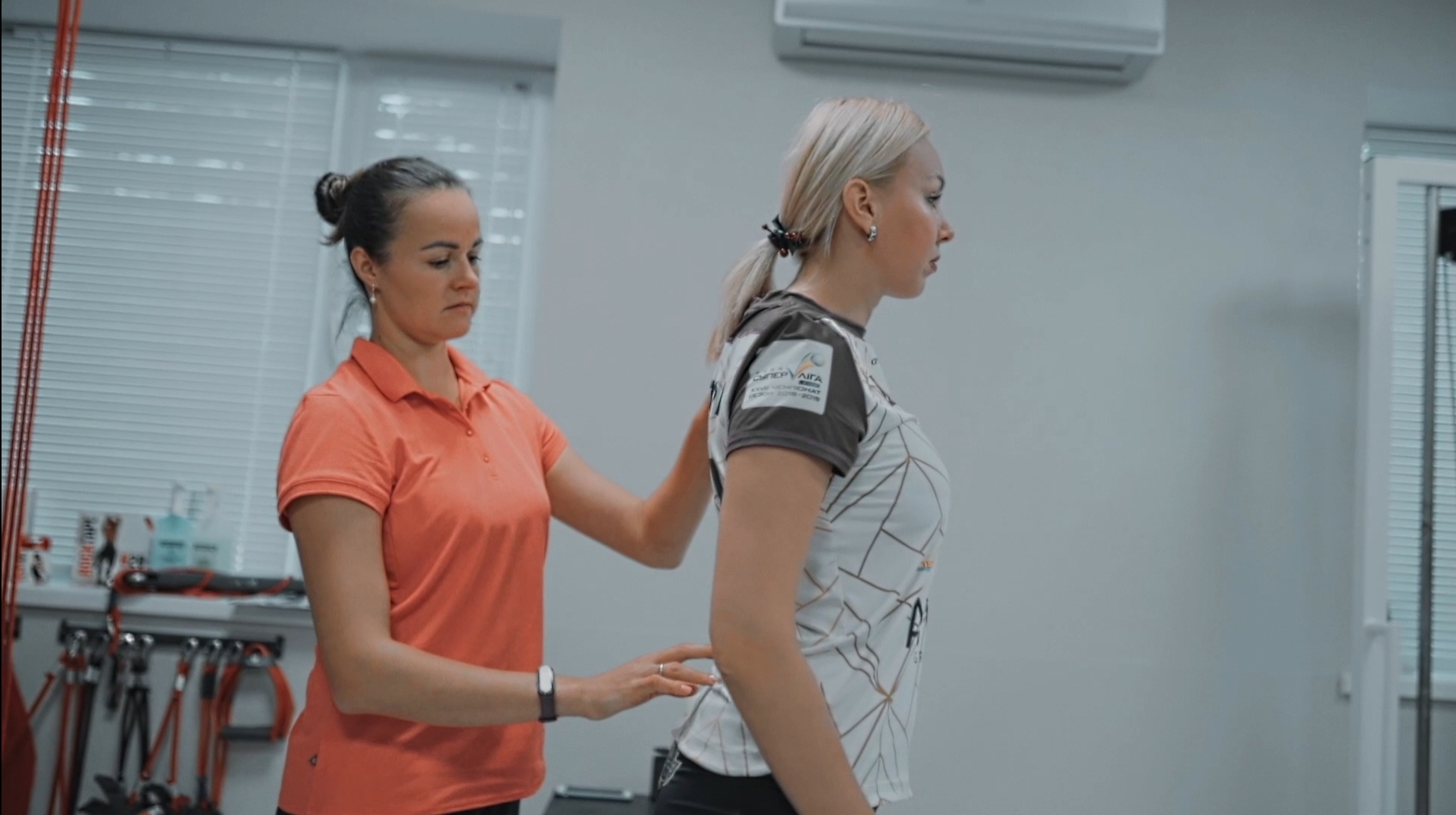
The specialist should correlate MRI results with your real physical symptoms. This includes functional tests: reflex assessment, muscle strength evaluation, nerve tension tests, and palpation of trigger points.
Step 3. Create a recovery plan based on physical rehabilitation
If neurological deficits (such as loss of reflexes or progressive muscle weakness) are absent, a conservative approach is the best option. The main focus should be on active restoration of musculoskeletal function.
How properly selected therapeutic exercises help the spine “heal”
Professional physical rehabilitation is based on the principle of controlled movement. Specifically selected therapeutic exercises address several critical tasks at once:
- Improving disc trophism (nutrition). Intervertebral discs in adults do not have their own blood supply. They receive water and nutrients exclusively through diffusion—like a sponge absorbing fluid during compression and decompression through proper movement.
- Stimulating hernia resorption. Moderate physical activity activates immune cells (macrophages). They recognize extruded disc tissue as foreign material and gradually break it down and reduce its size.
- Creating muscular protection. Since the affected spinal segment temporarily loses stability, surrounding muscles learn to compensate, reducing load on the injured area.
A competent physiotherapist will never offer a generic online exercise set. Exercises must be selected individually depending on the direction of disc protrusion and your functional capabilities.
FAQ
Do I need to repeat MRI every month to monitor my back?
No, doing so without a clear medical indication is unnecessary. Structural changes and disc resorption occur slowly. Repeat imaging is usually recommended no earlier than 6–12 months, or in case of sudden symptom worsening.
If protrusions are found on MRI, can I still exercise?
A protrusion is not a contraindication for physical activity. On the contrary, complete inactivity leads to deconditioning, weak core muscles, and faster disc degeneration. Only heavy axial loading and sudden twisting movements should be temporarily avoided.
Can a physiotherapist help if the hernia is large (e.g., 9–10 mm)?
Yes, the size of a hernia on imaging does not always determine treatment strategy. Large but soft extrusions often resolve faster than small but dense, long-standing protrusions. The key factor is symptoms and neurological function, not millimeters on a report.