



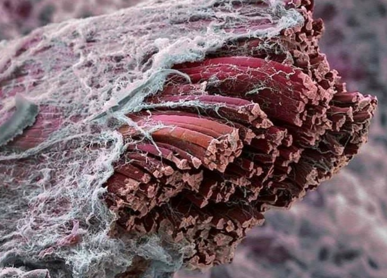
Fascia: The Mysterious Connective Tissue and Its Role in Our Bodies
Modern Definition and Role of Fascia:
Fascia is a continuous network of connective tissue that permeates our entire body. It is formed mainly by collagen fibers and is located under the skin, covering muscles, bones, internal organs and nerves with a single “cover” (Anatomy, Fascia Layers – StatPearls – NCBI Bookshelf). (photo 1)
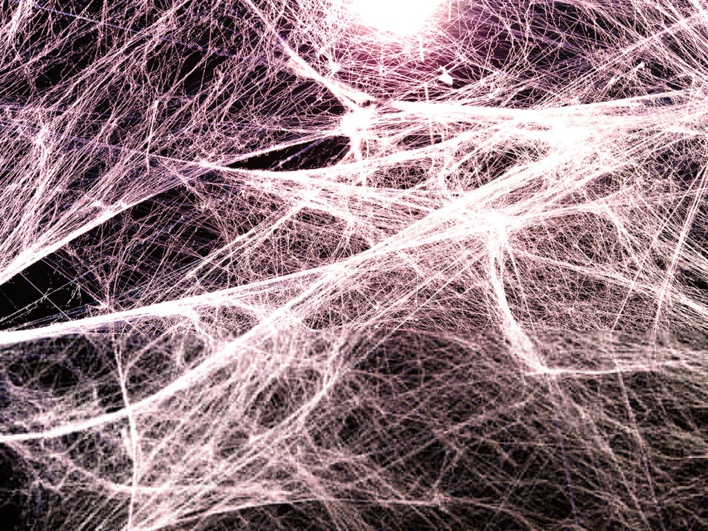
photo 1
In other words, fascia can be imagined as a kind of elastic overalls or a spider web inside us, which stretches continuously from head to toe. If you have ever cooked meat, you could see a thin white film on it – this is fascial tissue. Its main task is to connect and support the structures of the body: fascia envelops each muscle and organ, separates them from each other, while simultaneously fixing them in place, and also connects them together, forming a single whole. Through fascia, the force of muscle contraction is transmitted throughout the body; it holds organs in place and forms a framework for blood vessels and nerves (Fig. 1) (Anatomy, Fascia Layers – StatPearls – NCBI Bookshelf).
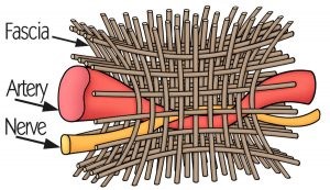
Fig. 1
Although fascia was previously considered simply a “packaging material” for muscles, new research reveals its active role. Fascial membranes contain a lot of elastin and fluid, which provides them with flexibility and low friction – thanks to this, muscles can slide freely relative to each other (Fascia (anatomy) – Wikipedia).
It is also important that the fascia is permeated with a network of nerve endings – both sensitive (sensory) and autonomic. Therefore, fascia is now considered an important sensory organ: it participates in the perception of the body’s position in space (proprioception) and the perception of pain (Fascia (anatomy) – Wikipedia). In other words, fascia is not just a passive membrane, but a dynamic system of perception and response that supports our posture, coordination of movements and general body sensation (photo 2). There is even evidence that fascia contains special cells (myofibroblasts) that are able to contract like smooth muscles under the influence of nerve signals – this confirms that fascial tissue can actively change its tone depending on the state of the body (for example, during stress).
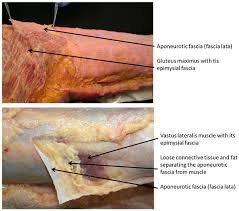
photo 2
Historical overview: from “forgotten” tissue to object of research
Historically, views on fascia have taken an interesting path. In old anatomical atlases, fascia
was often depicted, but little discussed – it was dissected to get to “more important” structures, such as muscles or organs. For many decades, fascia was considered an inert wrapper: surgeons and anatomists did not have a common opinion on what exactly to include in this concept. It is not without reason that the official anatomical nomenclature at the end of the 20th century noted that there is no single comprehensive definition of fascia, and different authors interpret it in their own way (Fascia – Wikipedia). In other words, there was no clear idea where “fascia” ended and other connective structures began. (Fig. 2)
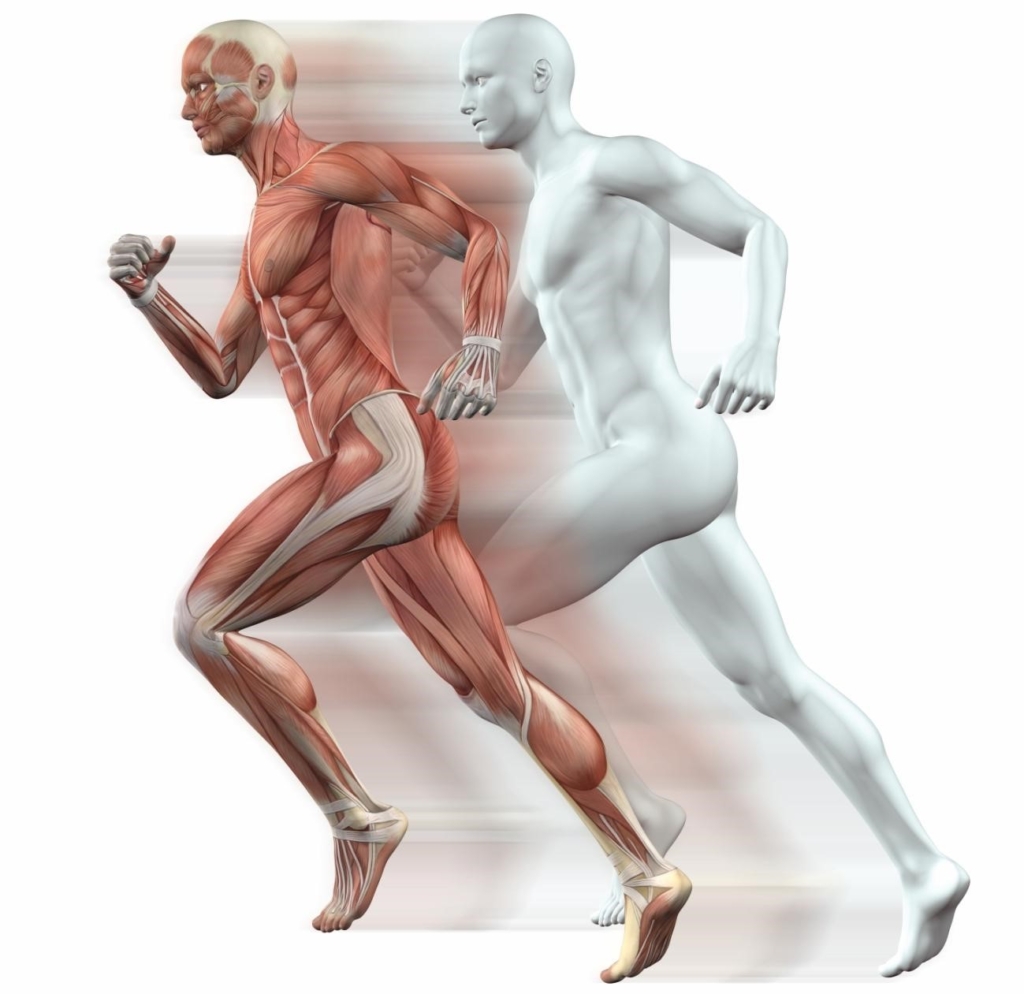
Fig. 2
Because of such underestimation, this tissue was even dubbed “forgotten”. It is noteworthy that in 2011 a scientific article appeared with the telling title “Fascia: The Forgotten Structure” (Anatomy, Fascia Layers – StatPearls – NCBI Bookshelf). In it and similar works, researchers called for attention to fascia as an important anatomical element.
The turning point was the end of the 20th and the beginning of the 21st centuries: osteopathy and manual therapy were developing, where fascia was given a key role, and new imaging methods (ultrasound, MRI) made it possible to see fascial layers in vivo. In 2007, the first International Congress on Fascia Research was held, which united scientists from different fields and caused an explosion of interest in this topic.
Over the past 10–15 years, fascia has moved from the shadows to the foreground: it began to be called an “organ” or “system” because of its integrity throughout the body, and the World Federation of Anatomists proposed more precise terms for the fascial system. Thus, from an almost ignored structure, fascia has become a hot topic in science, with dozens of studies published every year. Now no one doubts that without understanding fascia, it is impossible to explain many aspects of health and diseases of the musculoskeletal system.
The influence of various factors on fascia
The condition of fascia tissue is influenced by both external and internal factors. Fascia reacts to our lives: from the level of activity to stress and injuries. Let’s consider the main factors and the changes they cause:
Stress and muscle tension: Psycho-emotional stress often responds physically – we instinctively tighten our muscles (for example, our shoulders rise, our back tenses). Fascia is closely connected to the muscles, so chronic stress leads to chronic tension of the fascial network.
In a state of constant “combat readiness”, fascia loses elasticity, becomes less elastic. You can imagine that your inner thin, all-pervading frame begins to shrink and contract. This, in turn, reduces the flow of blood and nutrition to the tissues. Painful seals can form in the fascial layers – the so-called trigger points (Fig. 3)
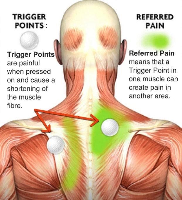
These changes often underlie myofascial pain syndrome, when a person experiences muscle-fascial pain and stiffness without obvious trauma. These phenomena are well known: who has not felt a “stone” in the neck after a stressful day? Interestingly, biologists have discovered: stress hormones (for example, adrenaline) can affect fascia cells (myofibroblasts), causing them to contract and further increase tissue tone. Thus, stress triggers a vicious cycle – tense fascia limits movement, and stiffness in the body adds discomfort and new stress.
Injuries, damage and inflammation: Any physical trauma – a sprain, a bruise or a surgical operation – disrupts the integrity of the fascial network. In response, the body activates inflammation and healing, during which fibroblasts form new collagen. Unfortunately, scar tissue is not as elastic and orderly as the original fascia (Fig. 4). Collagen fibers in the scar lie chaotically and “glue” together the layers that previously slid freely. As a result, adhesion is formed, a connective tissue “framework” that tightens the adjacent tissues. That is why after operations or deep wounds, a feeling of tightness often appears: the scar seems to “stick” to the muscles or internal organs.
The fascia in the area of injury thickens, loses elasticity – this is a manifestation of fibrosis (growth of healthy functional tissues with non-functional tight connective tissue). For example, after spraining the ligaments of the ankle joint, a person may still feel stiffness for a long time due to changes in the surrounding fascia.
In the worst case, persistent contractures (restriction of mobility) develop – a vivid example can be seen in severe burns, when scars on the skin and fascia reduce the volume of movements in the joints.
Scientific observations confirm: injuries and inflammation cause the formation of fibrous adhesions in the fascia, especially after surgical interventions and during healing, with the formation of scars (Fascia (anatomy) – Wikipedia). Thus, mechanical damage to fascia often has long-term consequences – the structure of the tissue changes, and this can leave a person with a legacy in the form of chronic stiffness or pain.
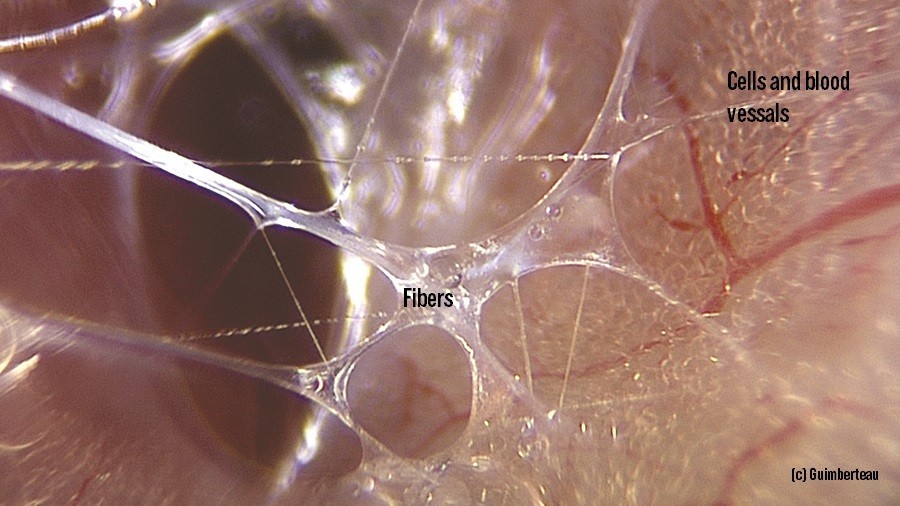
photo 3
Inactivity (hypodynamia): Our fascia needs movement, like water for a plant (photo 3). During normal activity, the collagen fibers of the fascia are constantly slightly stretched and displaced relative to each other, and fluid circulates between them, moisturizing the tissue. If a person leads a sedentary lifestyle, the fascia “drys out” and thickens. Imagine that you have kept rubber in improper conditions for a long time – it becomes hard and cracks when pulled.
Similarly, immobile fascia loses flexibility. In the absence of regular stretching, collagen fibers begin to stick together between layers (microadhesions are formed), hyaluronic acid in the intercellular substance thickens, and sliding weakens. Over time, the fascia can “dry out” and contract, limiting the normal amplitude of movements. It is not without reason that after a long stay without movement (for example, in a cast or simply at the computer) we feel that the body has frozen.
Inactivity also contributes to chronic pain: the stiff back of an office worker is partly associated with changes in the fascial structures of the back. Fortunately, these processes are reversible – it is worth resuming regular exercises, and the fascia will gradually restore most of its properties. But neglected hypodynamia can lead to a whole range of problems of the musculoskeletal system: from constant stiffness to an increased risk of injuries, because “untrained” fascia tears more easily. In other words, there is no movement – the fascia “rusts”.
Age-related changes: With age, all connective tissues undergo changes, and fascia is no exception. After the peak of physical fitness in youth, the body gradually decreases the content of water and elastic fibers, but the density of collagen increases. The fascia of an older person is stiffer and thicker, with more cross-links between fibers (like “knots” on threads). These structures hold the collagen together, but make it less flexible. As a result, an older person feels that their muscles and joints are “stiffening.” This is partly due to age-related changes in the fascia – they lose elasticity and cease to provide smooth tissue movement.
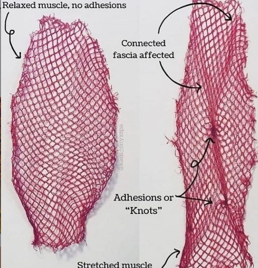
photo 4
In addition, the ability to recover also decreases: microtraumas of the fascia, which we constantly experience in life, heal quickly and without consequences in youth, but in old age they can accumulate, adding fibrous nodules and adhesions. Thus, the functionality of the fascia decreases with age – it absorbs loads worse, recovers more slowly after sprains, and supports muscles less elastically (photo 4). This is one of the reasons why stiffness and chronic pain in the back or joints often appear in the elderly. On the other hand, moderate physical activity can significantly slow down these age-related changes – we will talk about this later.
In general, various negative factors can lead to two types of fascia problems: either the tissue becomes too stiff, loses elasticity and “clamps” the muscles (as in myofascial syndrome), or vice versa – too relaxed and weak, loses its supporting function (as in some genetic hypermobility syndromes).
Both conditions are undesirable. Health lies in balance: the fascia must be elastic enough to support the organs, but also elastic enough not to restrict movement. Violations of the properties of fascia are a sign of pathology or a consequence of injury (Fascia (anatomy) – Wikipedia). That is why it is so important to understand how different factors affect this unique tissue and what we can do to maintain its optimal condition.
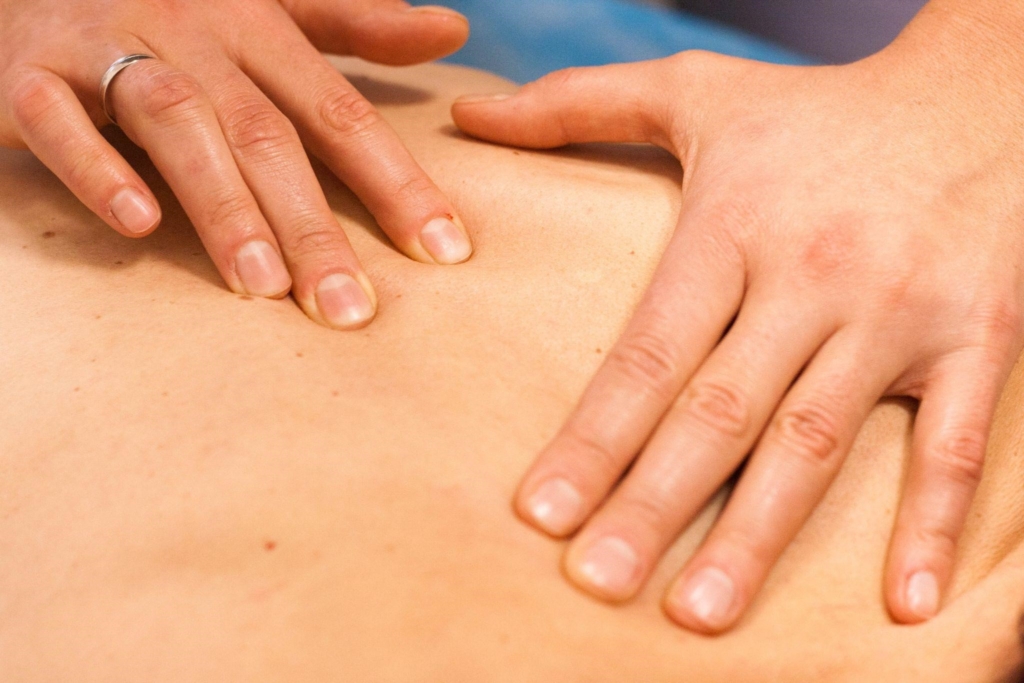
photo 5
Modern methods of influencing fascial tissue
Understanding the role of fascia in the body has contributed to the development of methods aimed at its improvement. Today, there is a whole arsenal of approaches – from special massage to physical exercises – that help improve the condition of fascia (Fascia (anatomy) – Wikipedia). Let’s consider the main ones and what science knows about them:
Manual techniques (myofascial massage, manual therapy): These are methods when the therapist affects the fascia with his hands – stretches, pushes, shifts layers of tissue (photo 5). An example is myofascial release: the specialist slowly and deeply passes through the soft tissues, as if “ungluing” the stuck fascia. The goal is to restore normal sliding between the layers, relieve excessive tension and thereby reduce pain and stiffness.
Many are familiar with the massage technique, when the skin and muscles are stretched in different directions – this is the effect on the fascia. Does it work? Studies in recent years have yielded encouraging results. A 2015 review of 19 clinical trials of myofascial release concluded that while the quality of the studies varied, the method generally shows a positive effect and is gaining a growing evidence base (Effectiveness of myofascial release: systematic review of randomized controlled trials – PubMed).
Patients with chronic back pain, fibromyalgia, and other conditions report relief after a course of this treatment. Manual techniques improve local blood circulation, stretch, and align the collagen fibers of the fascia. Interestingly, during such exposure, fibroblast cells react and begin to rebuild the extracellular matrix – in fact, manual therapy can trigger repair processes in the connective tissue. Thus, professional fascial massage is not only a pleasant rubbing, but also a serious recovery tool, the effectiveness of which is confirmed by scientific data.
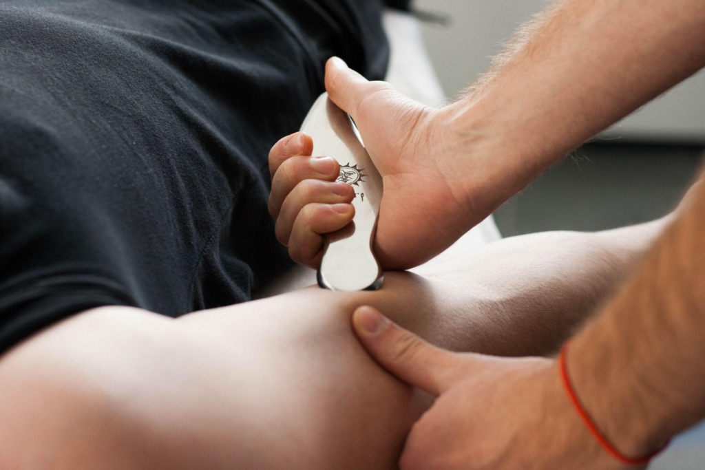
photo 6
Massage “blades” and instrumental techniques: In addition to hands, special tools are also used in fascial therapy. For example, massage blades are smooth metal or plastic plates that are used to “scrape” the skin. Graston, Gua Sha, and other types of IASTM (Instrument Assisted Soft Tissue Mobilization) techniques are popular in sports rehabilitation (photo 6).
The principle is similar to manual massage, but the blades allow for a more targeted and forceful impact on dense fascial seals and scars. They seem to “cut” adhesions in the tissue, improve microcirculation, and stimulate fibroblast renewal.
Scientific results: A 2019 systematic review showed that such techniques do improve joint mobility (ROM) in healthy people, and in patients with injuries – reduce pain and improve function of the affected area (Instrument-Assisted Soft Tissue Mobilization: A Systematic Review and Effect-Size Analysis – PubMed). In particular, it was noted that after the use of IASTM, muscle flexibility increased in athletes, and in patients with tendonitis, pain-free movement was restored faster.
In other words, the “scraping” massage tool is not a charlatan, but an effective technology: it works like a micro-“plow” that loosens hardened fascia. Of course, it is important that the procedure is performed by a trained specialist to avoid unnecessary trauma. When used correctly, instrumental techniques complement manual therapy and accelerate recovery from fascial injuries.
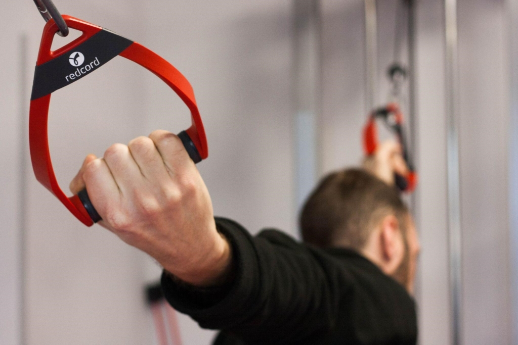
photo 7
Physical therapy and movement techniques: Fascia responds best to movement – so a variety of exercises are the key to its health. There are entire areas called “fascial training” or kinesitherapy, which focus on stretching and strengthening connective tissue structures (photo 7).
One simple and effective approach is regular stretching. Slow and gradual stretching of the muscles also provides a stretch for the fascia that covers them. It is important to stay in the stretched position for at least 30 seconds, or even 1–2 minutes – fascial fibers react more slowly than muscle fibers, and it takes time for them to give in. You may have noticed: the first few seconds it pulls hard, but then it seems to “let go” – this is the moment when the fascia begins to deform pliably. Such exercises increase the elasticity of the tissues and increase the amplitude of movements.
Another component is dynamic movements and vibrations. For example, jumping, bouncing, wave-like exercises (like in tai chi) train the elasticity of the fascia. There are even special rollers and balls for myofascial self-massage (foam rolling): a person rolls over problem areas with his own weight, kneading the fascia – this helps to relieve muscle tension and improve flexibility.
Scientific experiments have confirmed that a combination of stretching and moderate physical exertion improves the condition of the fascia tissue. Physical activity stimulates fluid exchange: when muscles contract and stretch, the fascia is squeezed and then saturated with moisture again, like a sponge – this prevents it from drying out and sticking together. In addition, the loads contribute to the production of collagen in the right places and directions: fibroblasts orient new fibers along the lines of tension, which increases the strength of the tissue.
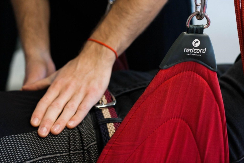
photo 8
Thus, properly selected exercises can “re-educate” even damaged fascia, returning its flexibility and functionality (photo 8). Of course, physical therapy does not give an instant effect – it is a gradual process that requires regularity. But the plus is that a person is actively involved in his recovery. For example, after joint operations, patients are prescribed a set of movements to prevent the formation of adhesions – and this is largely the task of working with fascia. In general, active movement is one of the most effective “tools” for fascia that we have.
Usually, the best result is given by a combination of methods. First, say, manually or instrumentally develop overly hardened areas (to “unblock” the fascia), and then consolidate the effect with exercises so that the tissue adapts to a healthy state. In the arsenal of specialists there are other auxiliary means: heat to increase the elasticity of the fascia, water procedures and even electric stimulation – everything that can improve blood circulation and relieve tension indirectly helps the fascia.
If conservative methods do not give results, sometimes they resort to surgical intervention – for example, in case of pinching syndromes, fascial partitions are cut open to free nerves or muscles. But this is an extreme case. In most situations, fascial dysfunctions can be treated without a scalpel – all that is needed is time and the right approach. The main thing that modern medicine recognizes: it is necessary to work with fascia (Fascia (anatomy) – Wikipedia), otherwise the treatment of many chronic pains or recovery from injuries will be incomplete.
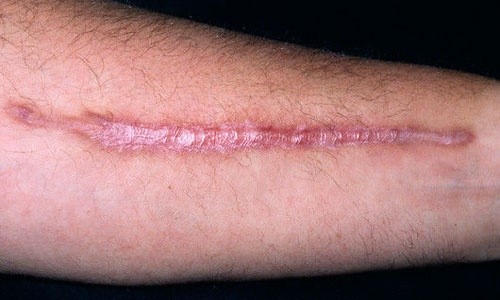
photo 9
Scars and adhesions: how scars limit mobility
It is worth talking about scars separately – after all, almost everyone has at least a small scar, and many have encountered postoperative scars. Scar tissue is a natural result of the healing of deep wounds, but it can become a source of problems. As we mentioned, a scar on or near the fascia often leads to adhesion – adhesions between the fascia and neighboring tissues (Fascia (anatomy) – Wikipedia) (photo 9).
Why does this happen? During healing, fibroblasts fill the damaged area with collagen, forming a dense knot. This “patch” connects the edges of the wound, but also sticks and grows to everything around it. Normally, our internal structures should move slightly relative to each other (for example, when raising an arm, muscles slide under the skin). If adhesions have formed, movement causes pulling discomfort: the skin seems to be attached to deeper layers. Sometimes, the scar pulls even distant areas – for example, after abdominal surgery, patients feel pulling and tugging in the pelvic area during various movements. This is explained by the fact that the fascial network is connected throughout the body, and a large scar disrupts the tension of the “tissue web” more widely than it seems.
Consequences of scar adhesion to fascia:
- Limited mobility. Joints do not reach the full range of motion, muscles do not stretch to the end. Hence the feeling of stiffness, a foreign body or even pain when trying to make a certain movement.
- Circulation disorders: adhesions can compress small blood vessels or lymphatic vessels, which leads to poorer tissue nutrition and swelling.
- Neurological symptoms: if the scar presses on a nerve, tingling, numbness or shooting pains are possible. For example, after a cesarean section, numbness sometimes occurs around the scar – this is a sign of the involvement of nerve endings in the scarring process.
What to do with scars? Fortunately, fascia is a living tissue, and even after the formation of a scar, it is gradually rebuilt. Proper rehabilitation can significantly reduce the negative effects. Scar massage (manual or vacuum) is practiced – it improves the elasticity of scar tissue and prevents it from fusing with the surrounding area. Stretching and movement are also effective: gentle, regular stretching of the scar area stimulates collagen remodeling so that the fibers align along the lines of movement.
If you start rehabilitation on time (even while the wound is still healing), you can prevent the formation of rough adhesions. Old scars can also be corrected, although more slowly – sometimes a combination of physiotherapy and even injections of enzymes that soften fibrosis are used.
It is important to understand that scar tissue is less elastic than normal fascia, so it will not become completely “like new”, but competent treatment can significantly improve the situation. As a result, although the scar remains forever, it should not “tighten” your life – modern medicine has methods that return maximum mobility to the fascia around the scar.
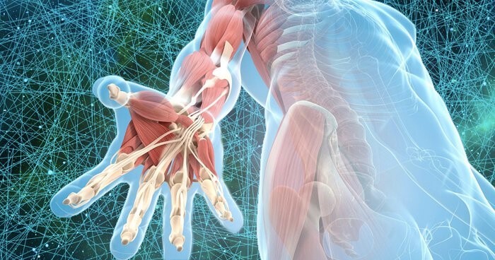
Fig. 5
Physical activity – support and restoration of fascia
As we have already mentioned more than once, movement is life for fascia (Fig. 5). Physical activity plays a key role in maintaining the normal structure of connective tissue and even in its regeneration after injuries. An analogy is appropriate here: fascia is like an elastic plastic lattice, which is stronger and healthier the more it is moderately stretched and compressed. Without a load, the lattice is fragile, and with excessive load, it tears. So, balance and regularity are needed.
For the prevention and maintenance of fascia in tone, moderate dynamic loads are best suited. Regular walking, jogging, swimming, yoga – all these types of activity force the fascia to work in different modes, which maintains its elasticity. The WHO recommendations for 150 minutes of moderate activity per week are also useful for connective tissues. Alternating loads – for example, strength exercises and stretching – provides the fascia with optimal stimuli: during strength work, collagen fibers receive microdamage, and during rest and stretching they are arranged more orderly and firmly.
It is important to pay attention to flexibility: at least 2-3 times a week to do stretching complexes or do yoga/Pilates. Studies show that after 4-6 weeks of regular classes, flexibility increases noticeably, and the feeling of stiffness in the body decreases – largely due to positive changes in the fascia. Period is no less important: it is better to move a little every day (for example, get up and stretch every hour if you sit at work) than to exhaust yourself in the gym once a week. Micro-movements throughout the day are useful for fascial tissue – stretching in different directions, making circular movements with your arms, bending backwards, etc. This supports fluid circulation and prevents fibers from sticking together.
For a therapeutic effect with existing fascial problems, physical activity is used in a more dosed and targeted manner. For example, after an injury, a period of immobilization (rest) is required, but as soon as the acute stage has passed, dosed movements should be introduced in the area of injury. At first, these are passive movements with the help of a rehabilitation specialist, then – active exercises without load, and later – strengthening. The goal is to direct fascial remodeling in the right direction. If this is not done, the fascia will recover chaotically (with the formation of scars and adhesions).
So, rehabilitation exercises are a kind of “instructions” for fibroblasts on how to rebuild tissue. It is important to adhere to the principle of gradualness: the intensity and duration of classes are increased step by step so as not to overload the fragile new fibers. Therapeutic physical education usually lasts months – but as a result, the fascia returns to the maximum possible normal state.
What intensity and duration of movement is needed? Of course, this depends on individual characteristics and health status. But in general, to maintain the fascia, moderate aerobic activity for at least 30 minutes a day (or 3–4 times a week) plus 10–15 minutes of stretching for the main muscle groups is recommended.
Moderate activity is when you feel a little short of breath, but you can talk (for example, fast walking, light running). It is with such a load that blood circulation in the small vessels of the fascia improves, and the muscles work without tearing, which contributes to the healing of the connective tissue. Gentle stretches are often used to treat fascial pain, with fixation for 1–2 minutes and repetition several times a day – this allows you to gradually restore mobility in a few weeks.
It is also important not to overdo it: excessively intense training without sufficient recovery can, on the contrary, injure the fascia. For example, marathon running without preparation is a great stress for the connective tissues, it can cause micro-tears of the plantar fascia or other structures. Therefore, the therapeutic effect is provided by regular, but dosed load. It is better to do it longer and slower than quickly and too hard.
Finally, let’s mention another very important factor – hydration. Fascia is 60–70% water (in the intercellular substance), so the drinking regime is also important: dehydration and deficiency of trace elements make it stiffer. Active people usually drink more themselves, which has a good effect on the condition of the tissues.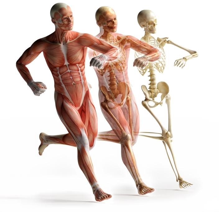
Fig. 6
Conclusion
Fascia has evolved from an inconspicuous connective tissue into a real star of modern anatomy. Today we understand that the fascial system is a holistic organ that encompasses our entire body, forming a single framework and environment for muscles, organs, blood vessels, and nerves. Fascia is unique in that it combines seemingly contradictory properties: it is both strong and elastic; it is capable of transmitting mechanical forces and sensing the most delicate changes (pressure, pain); it can both relax and contract. It is not for nothing that it is called the organ of form – it is the fascial “suit” that gives our body integrity and shape.
The importance of fascia for health cannot be overestimated. Our flexibility, posture, coordination of movements, and the absence of pain during movements depend on the condition of this tissue. A “failure” in the fascial system can manifest itself in various problems – from a stiff back and headache (due to tension in the tendon helmet on the skull) to a limitation in raising the arm (due to a postoperative scar). Fortunately, fascia is a very plastic and trainable structure. It is constantly rebuilt throughout life, so we have the opportunity to influence it.
Regular physical activity, reasonable therapy methods and self-care can maintain fascia in excellent condition until old age. Conversely, neglect of movement or chronic stress can quickly “outdated” our fascial system, causing pain and stiffness.
The good news is that medicine now has tools to work with fascia. Chiropractors, physiotherapists, osteopaths – they all actually deal with the fascial system, even if they didn’t call it that before. New studies confirm the effectiveness of many approaches, which means that patients can count on real help.
Of course, there are no miracles: it is impossible to deal with chronic “clamps” or old adhesions in one session. Fascia needs time to change. But, given its pervasiveness, even small improvements can give a tangible result – after all, when the fascia is “released”, a feeling of lightness and freedom of movement spreads throughout the body.
In conclusion, we can say that fascia is a kind of “silent ascetic” of our body. It imperceptibly performs a colossal job, supporting us every moment. Now that science has lifted the curtain on its secrets, we have a chance to help this hero.
Let’s take care of our fascial system – let’s move, avoid chronic overstrain, treat injuries in a timely manner. And then our mysterious internal web will serve us reliably and imperceptibly – as befits a true hero, whose existence we may even forget in everyday life, but whose importance cannot be overestimated.
Our team takes a comprehensive approach to solving various issues of restoring movement after injuries, fractures, and surgeries. We combine various techniques, methods, and approaches in order to restore our clients as quickly and qualitatively as possible.
If you need help to quickly and qualitatively return to an active motor life, contact the specialists of our PhysioSpace functional recovery center in Irpin at V. Stus, 27., tel. 0937001040.