



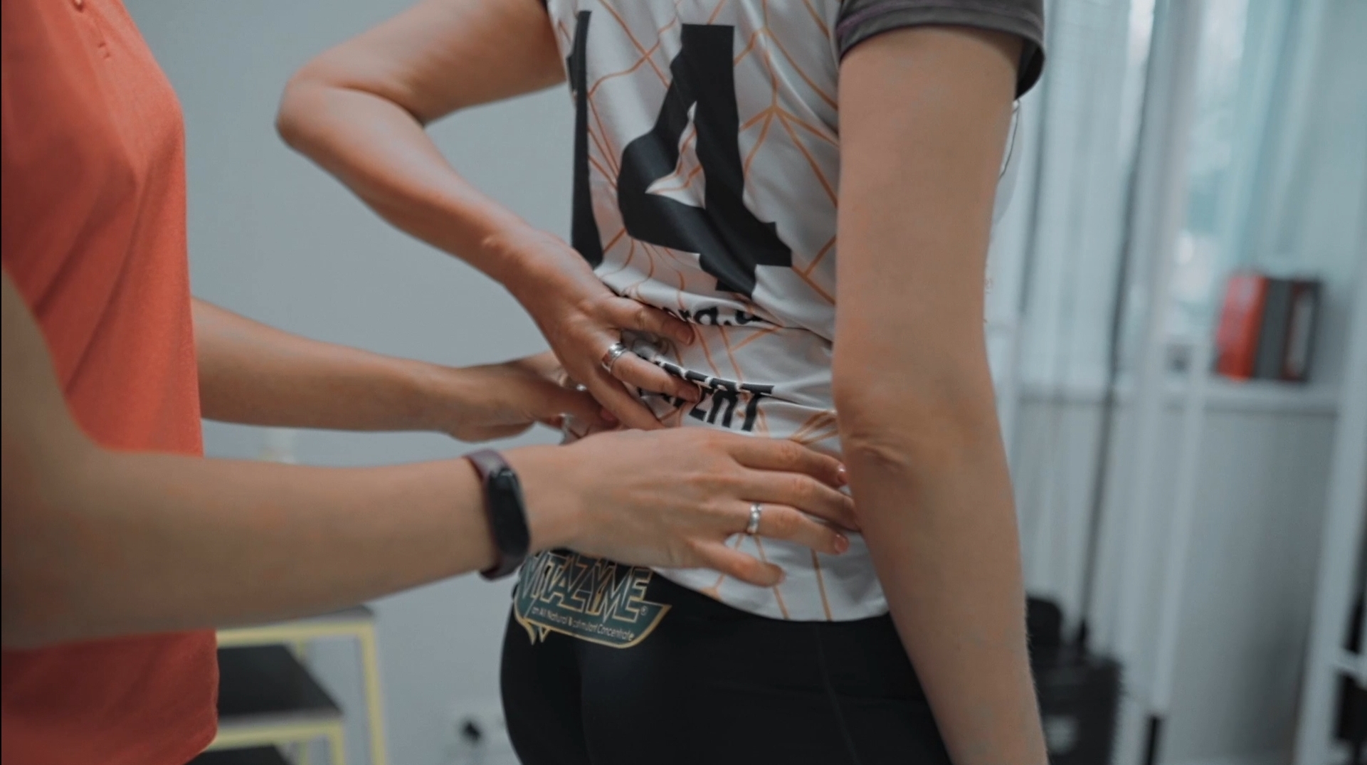
A sudden sharp “electric” pain in the lower back that radiates down the leg to the toes, or numbness in the arm after prolonged computer work, often causes panic. In everyday language, people tend to label this kind of symptom as a so-called “pinched nerve.” They immediately seek manual therapists to “put the vertebrae back in place” or start taking strong painkillers uncontrollably.
However, in the official International Classification of Diseases, such a diagnosis simply does not exist. What people call a “pinched nerve” is actually a complex neurochemical and mechanical process that requires a completely different rehabilitation approach. In this article, we will break down the real anatomy of this condition, explain how neuropathic pain develops, and why aggressive physical manipulation can seriously harm the nervous system.
The common myth: why you cannot simply “put back” a supposedly pinched nerve
The idea that two vertebrae can shift and trap a nerve root like a passenger in a subway door is a serious anatomical misconception. A spinal nerve root exits through relatively spacious bony openings. To mechanically compress it, the vertebrae would have to be severely damaged, such as in a major trauma or fracture.
Attempts to “adjust” the spine using forceful manipulation during acute pain are extremely dangerous. If the nerve tissue is already irritated, such aggressive interventions only increase protective muscle spasm, worsen local swelling, and may even lead to irreversible nerve damage.
The real anatomy of pain: compression and inflammation of the nerve root
So what actually causes this intense burning pain? In 90% of cases, the cause is radiculopathy (or radicular syndrome)—a pathological process involving both mechanical compression and chemical inflammation. When an intervertebral disc bulges, it may come into contact with the nerve sheath.
The contact itself is not always painful. However, the disc’s internal material contains specific proteins that are highly irritating to nerve tissue. When they come into contact, a strong chemical reaction occurs—local inflammation. The nerve swells, space becomes limited within the bony canal, its blood supply is disrupted, and the brain receives a signal of critical danger.
True nerve compression vs. muscle spasm: how to tell the difference?
People often confuse simple myofascial pain (trigger points) with true neurological injury. However, these conditions have key differences that determine the correct rehabilitation strategy.
| Comparison criterion | Radicular syndrome (radiculopathy) | Myofascial pain (muscle spasm) |
| Pain type | Sharp, burning, electric-shock-like, radiating. | Dull, aching, localized, felt as a deep tight band. |
| Pain distribution | Follows the nerve pathway (e.g., from buttock down the leg to the foot). | Localized to one muscle, with minimal referred pain. |
| Associated symptoms | Numbness, tingling (“pins and needles”), muscle weakness, reduced reflexes. | No sensory loss; strength and reflexes remain intact. |
| Effect of movement | Worsens with bending, coughing, sneezing, or spinal loading. | Worsens with stretching or prolonged static muscle load. |
(According to clinical guidelines of the European Academy of Neurology, true radiculopathy accounts for only 5–7% of all cases where patients report acute back pain. In the remaining 93% of cases, the source of pain is muscles, ligaments, and fascia.)
Main causes of true neuropathic pain
True irritation of nerve structures results from specific biomechanical and anatomical changes in the spine.
1. Herniated disc or disc protrusion
This is the most common cause. When the fibrous ring of the disc tears, the gel-like nucleus escapes and creates compression in the area where the nerve root passes.
2. Central or foraminal stenosis
Narrowing of the canals through which nerves pass. It usually develops in older adults due to bone overgrowth (osteophytes) or thickening of ligaments caused by chronic spinal wear.
3. Tunnel syndromes (peripheral compression)
In this case, the nerve is affected not at the spine but in peripheral narrow anatomical spaces (tunnels) formed by muscles and tendons. Examples include carpal tunnel syndrome in the wrist or piriformis syndrome in the gluteal region.
How evidence-based rehabilitation works for nerve injuries
Once acute chemical inflammation is managed with medication (prescribed by a physician), physical rehabilitation becomes essential. The goal of a physical therapist is not to mechanically “free” the nerve, but to create optimal conditions for its recovery.
Modern approaches include:
- Neurodynamics — gentle exercises that improve the mobility (gliding) of the nerve relative to surrounding muscles and tissues, reducing internal swelling and improving signal conduction.
- Decompression therapeutic exercises — positional movements that temporarily increase space around the nerve root, reducing mechanical pressure.
- Strengthening stabilizing muscles — rebuilding the deep muscular corset that protects the spine from micro-movements that irritate nerve tissue during walking or sitting.
FAQ
How long does a damaged nerve take to recover?
Nerve tissue regenerates slowly—about 1 mm per day after decompression is relieved. Therefore, treatment and rehabilitation of radiculopathy may take from several weeks to 3–4 months.
What if my leg is getting weaker or my foot is “dropping” due to back pain?
This is a critical symptom indicating serious impairment of motor nerve conduction. In such cases, you should immediately consult a neurologist or neurosurgeon, as prolonged weakness may lead to irreversible muscle atrophy.
Is surgery always required for a herniated disc compressing a nerve?
In 90–95% of cases, surgery is not required. The body is capable of lysis (natural reduction and resorption of the herniation) with proper conservative treatment and a well-structured physiotherapy program.